Child neurologists rely on clinical tests to accurately diagnose and treat their patients.
There are a number of reasons that a child neurologist might utilize a clinical test, including to make an initial diagnosis, for ongoing monitoring of the development of neurological conditions after a diagnosis, and to gauge the progress of treatments.
Here, we’ll get into the most common clinical tests that you might expect your child neurologist to order when you bring your son or daughter to the clinic.
Imaging Tests; MRI (magnetic resonance imaging) and CT Scans
Technology allows us to take a look inside of the brain, brain stem, and spine with a variety of tests. Tests that produce pictures of what’s going on in the brain are called imaging tests.
Imaging tests can identify a range of neurological conditions, such as:
- brain tumor
- infection
- inflammation
- traumatic brain injury (TBI)
- genetic conditions
- stroke
- multiple sclerosis
MRI (Magnetic Resonance Imaging) For Child Neurology
MRI combines the use of powerful (but safe) magnets and radio waves to generate highly detailed images of brain and nervous system tissue. They can also assess brain blood flow (important for optimal brain health) and detect mineral deposits in the brain.
There are two types of MRI:
- With contrast. In this test, the clinician injects a dye (gadolinium-based) into the patient’s veins, which then travel throughout the body into the brain. MRI with contrast produces clearer, more detailed images than MRI without contrast.
Patients with certain kidney conditions should not receive MRI with contrast. Rarely, the contrast dye may produce (usually mild) side effects like dizziness or shortness of breath. The body absorbs or eliminates the contrast dye shortly after the procedure. - Without contrast. MRI without contrast may be preferable in certain circumstances. Often, your doctor will determine that MRI without contrast is just as effective (in addition to being more affordable) and will therefore order this type of MRI.
Both MRI with and without contrast are effective imaging tests, but your provider may opt for one or the other based on certain factors like the reason for the test and the individual characteristics of the patient.
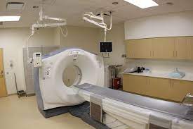
Computerized Tomography (CT Scans) For Child Neurology
A CT scan is a series of high-definition X-ray images taken from different angles. Taken together, these shots from different angles are combined into a three-dimensional profile of the brain.

CT scans in child neurology may be useful for:
- Identifying tumor location
- Finding a blood clot
- Diagnosing seizure disorders
- Assessing potential damage from traumatic brain injury (TBI) – for example, following a suspected concussion
CT scans are simple, quick, and usually do not require any sedation for your child. However, there is some concern among child neurologists regarding the risks of radiation exposure from CT scans:
“The rate of computed tomography (CT) scans in children has dropped in the last decade, with a move to radiation-free magnetic resonance imaging (MRI) scans and ultrasounds.”
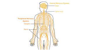
EEG (Electroencephalogram)
Child neurologists use EEGs to measure electrical signaling in the brain. They are largely risk-free and painless. Timewise, the tests usually take between 45 minutes and 2 hours. In some cases, your child might need a “prolonged” EEG test that takes 24 hours.
To perform EEG, the clinician places a series of small discs called electrodes over the brain and brain stem. The electrodes record the electrical activity and produce the results in graph form for interpretation by a trained expert.

Image source: Sebastian Nagel
EEG may be useful for diagnosing or assessing the extent of multiple neurological conditions in children, such as:
- Epilepsy
- Traumatic head injury
- Brain inflammation (encephalitis)
- Sleep disorders*
- Brain tumor
*If your child is undergoing testing for a sleep disorder, the test may be done overnight.
Lumbar Puncture (Sometimes Called Spinal Tap)
In a lumbar puncture, the clinician takes a sample of cerebrospinal fluid (CSF) from the spinal column (subarachnoid space) for lab analysis. The lab worker will analyze the CSF for:
- Its makeup of proteins, sugars, and white and red blood cells
- Its consistency and color
- The presence of pathogens like bacteria or viruses

The conditions that lumbar puncture can diagnose include:
- Bleeding in the subarachnoid space
- Reye syndrome
- Some types of cancers
- Encephalitis (brain swelling
- Meningitis (inflammation in the membrane coating the brain and spinal cord)
- Myelitis (spinal cord inflammation)
- Guillain-Barre syndrome
Because the needle punctures the spinal column, there is some risk associated with a lumbar puncture. For example, CSF leak can cause a headache that usually resolves shortly afterward. Also, there is a small risk of bleeding in the spinal canal after the procedure. Your doctor will balance the importance of the test with any potential risk factors.
Blood Tests
Your child neurologist might order a small blood sample from your child for lab analysis, usually taken from the arm. Blood tests are useful for diagnosing neurological conditions, for assessing their severity, and for monitoring the therapeutic effects of medications.
Blood tests can identify:
- Genetic conditions through DNA analysis
- Autoimmune conditions
- Bacterial or viral infections
- Blood clotting disorders
- Chronic inflammation
Positron Emission Tomography (PET)
PET scans assess metabolic function and the biochemical composition of brain tissue. The clinician injects radioactive isotopes called “tracers” which then produce 2D or 3D images for analysis. The type of tracer that your child neurologist uses will depend on the exact purpose of the test.
The PET scan procedure usually takes about 30 minutes to an hour.
PET scans perform multiple vital diagnostic functions:
- Diagram blood flow
- Detect tumors or diseased tissue
- Gauge cellular/tissue metabolism
The PET scanner is a large, open-ended, oval-shaped unit, similar in appearance to an MRI or CT scanner. Critically, PET scans focus on function rather than structure like other imaging tests – how your child’s body responds to various stimuli. They highlight areas of high metabolic and/or biochemical activity.
For example, the tracer fluorine-18 isotope is similar in structure to glucose (blood sugar) and is therefore useful for assessing how the body responds metabolically to the introduction of sugar into the bloodstream.
Contact Child Neurology Center of Northwest Florida
Whether you are interested in scheduling an appointment for your child or want to learn more about the tests available at our state-of-the-art clinic, please don’t hesitate to contact us – our friendly, knowledgeable staff is always ready to talk to concerned parents. Your child’s optimal health is our mission.